
Heart Anatomy and PhysiologyReview
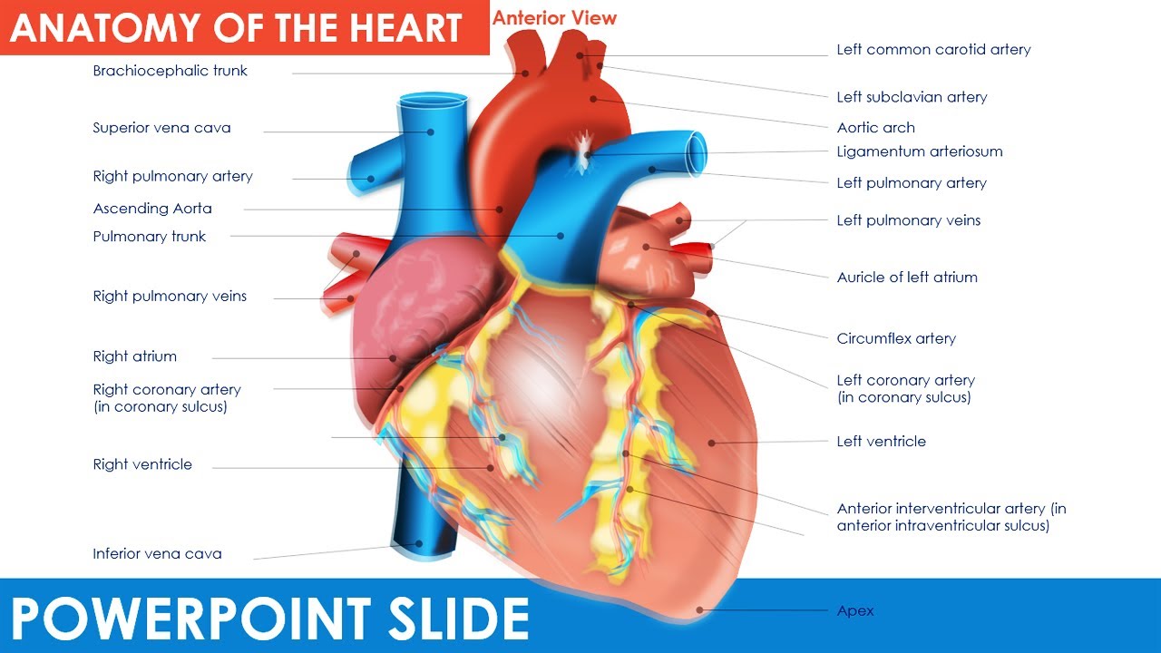
CPR. The position of the heart in the torso between the vertebrae and sternum (see Figure 19.2 for the position of the heart within the thorax) allows for individuals to apply an emergency technique known as cardiopulmonary resuscitation (CPR) if the heart of a patient should stop. By applying pressure with the flat portion of one hand on the sternum in the area between the line at T4 and T9.
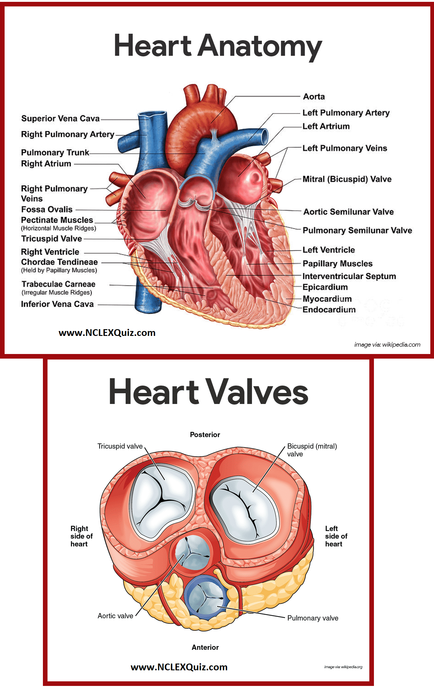
Diagram of Heart Blood Flow for Cardiac Nursing Students NCLEX Quiz
The heart is highly metabolically active and boasts the highest oxygen consumption by mass of any organ. This demand for oxygen is met by the coronary circulation, which is responsible for delivering blood to the myocardium and represents approximately 5% of cardiac output.[1] Adequate blood flow through the coronary vessels is critical to avoid ischemia and maintain the integrity of the.
PPT Anatomy and Physiology of the Heart PowerPoint Presentation ID7043704
Osmosis Cardiovascular Anatomy and Physiology high-yield notes offers clear overviews with striking illustrations, tables, and diagrams. Make learning more manageable.
PPT Heart Physiology PowerPoint Presentation, free download ID3130226
Anatomy and physiology of heart | PPT Anatomy and physiology of heart Dec 19, 2019 • 12 likes • 2,226 views Amrutha nayaka medical student Health & Medicine A small and clear introduction of Heart Anatomy and its function. Anatomy and physiology of heart 1 of 23 Download Now Save slide Save slide Recommended

Human Heart Diagram and Anatomy of the Heart StudyPK Heart diagram, Anatomy of the heart
Functions of the Heart The functions of the heart are as follows: Managing blood supply. Variations in the rate and force of heart contraction match blood flow to the changing metabolic needs of the tissues during rest, exercise, and changes in body position. Producing blood pressure.

Normal Anatomy of the Human Heart Giclee Print by Nucleus Medical Art at Chirurgia
Anatomy & Physiology: BIO 161 / 162. AP BIO 161 / 162; AP 1: BIO161 Toggle Dropdown. Chapter 1: An Introduction to the Human Body ;. Heart Anatomy. PowerPoint: Chapter 19, Heart Physiology. Outline: Chapter 19, Heart Physiology. AnatomyTV Coverings Activity. Cardiac Cycle Problem Solving. Mixed Up Blood Flow Pathway. Concept Map of the Heart.

PPT Anatomy and Physiology of the Heart PowerPoint Presentation ID7043704
The heart is a hollow, muscular organ about the size of a fist. It is responsible for pumping blood through the blood vessels by repeated, rhythmic contractions. The heart is composed of cardiac muscle, an involuntary muscle tissue that is found only within this organ. The term "cardiac" (as in cardiology) means "related to the heart" and comes
Anatomy of the Heart PowerPoint template Free Download PowerPoint University YouTube
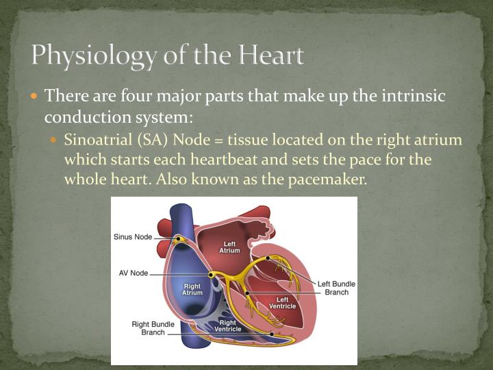
Anatomy, physiology and pharmacology of the autonomic supply of the heart. Anatomy, physiology and pharmacology of the autonomic supply of the heart. The Medulla control centre. The medulla is the primary site in the brain for regulating sympathetic and parasympathetic (vagal) outflow to the heart and blood vessels. 299 views • 13 slides
On Heart Kardiohirurgija.rs
The conducting system of heart by The conducting system of heart Idris Siddiqui 140.5K views • 26 slides Anatomy of heart by Anatomy of heart Dr. Binu Babu Nursing Lectures Incredibly Easy 158.4K views • 71 slides Cardiac conduction system by Cardiac conduction system Michael Wrock 107.1K views • 18 slides Heart Anatomy by
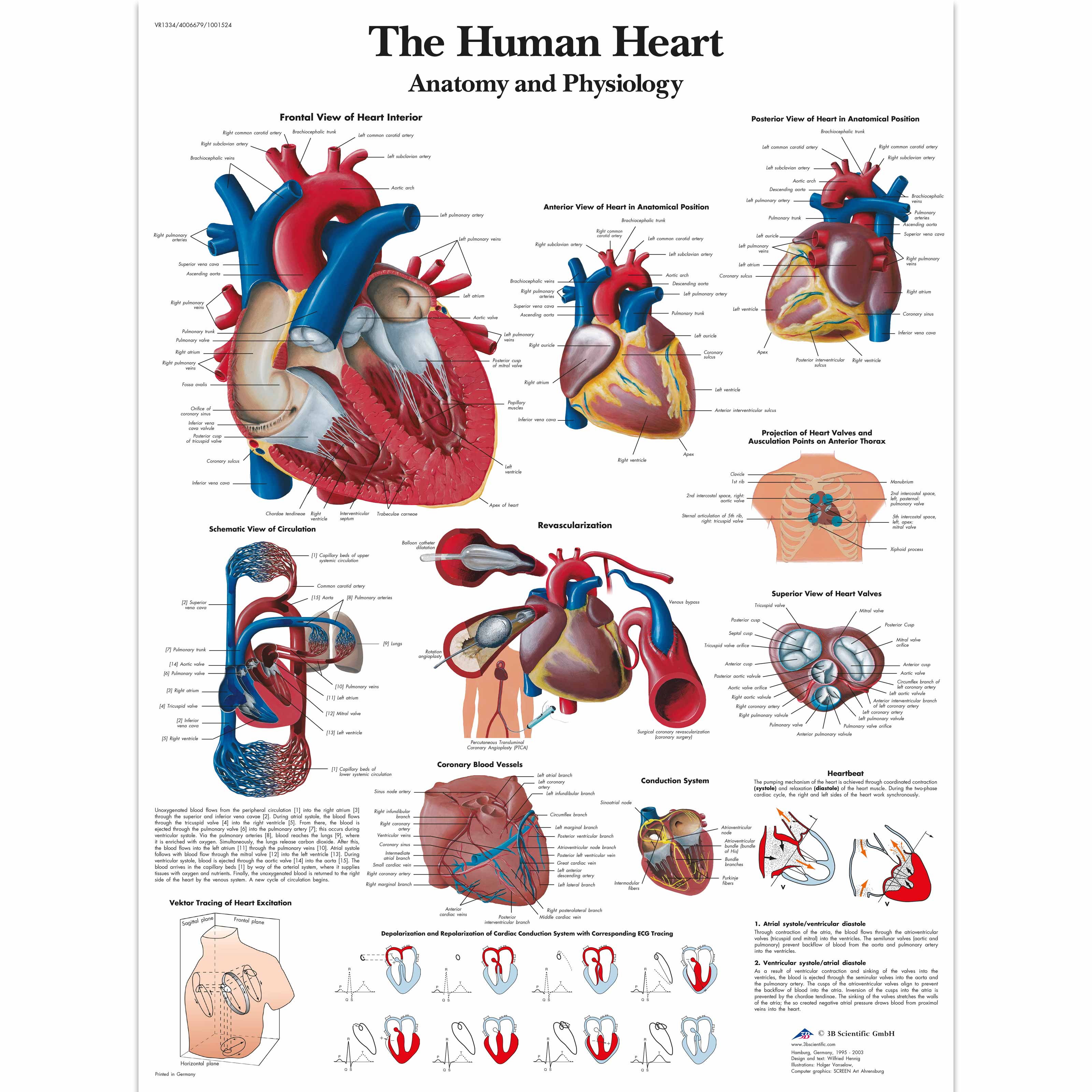
The Human Heart Chart Anatomy and Physiology 4006679 VR1334UU Cardiovascular System 3B
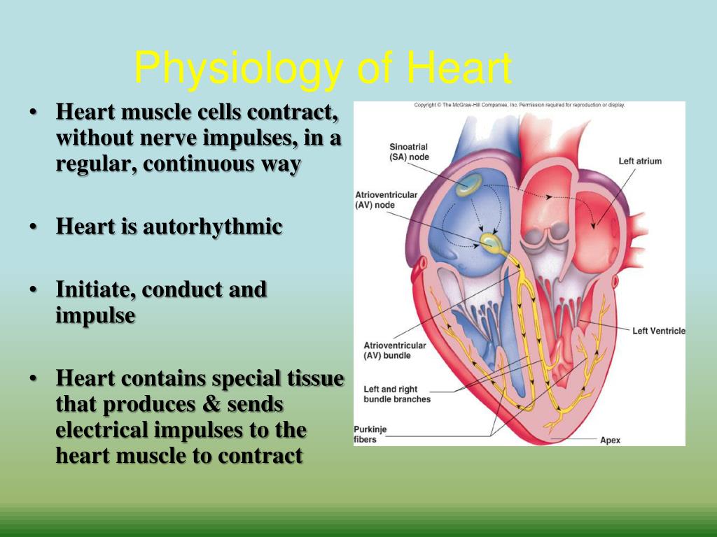
Cardiac physiology is one of healthcare's most important aspects of medical knowledge. The cardiovascular system constantly adapts to maintain homeostasis in the body, specifically to maintain oxygen perfusion of tissues. The heart will adapt via multiple variables such as heart rate, stroke volume, preload, afterload, diastole, and systole.

Human Heart Anatomy Diagram coordstudenti
3. Functions of the cardio vascular system: • Distribution of oxygen and nutrients to all the body parts • Transportation of CO2 and metabolic waste products from tissues to lungs and other excretory organs • Distribution of water electrolytes and hormones through out the body • Part of immune system • Thermoregulation. 4.

PPT Apparatus for the Analysis of Heart Sounds PowerPoint Presentation ID2963554
The heart is a muscular organ that serves to collect deoxygenated blood from all parts of the body, carries it to the lungs to be oxygenated and release carbon dioxide. Then, it transports the oxygenated blood from the lungs and distributes it to all the body parts. The heart pumps around 7,200 litres of blood in a day throughout the body.; The heart is situated at the centre of the chest and.

Heart Anatomy/Physiology
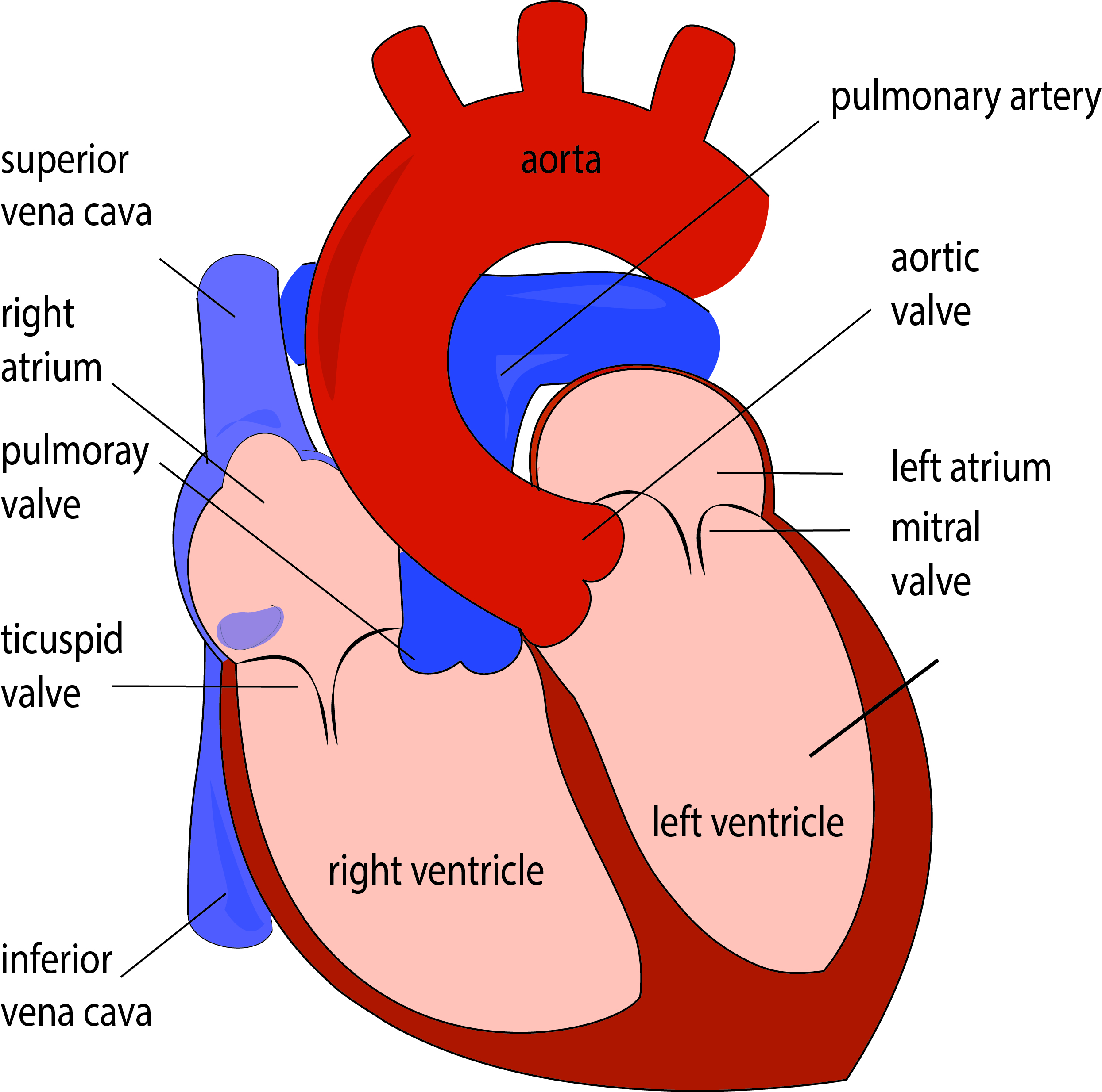
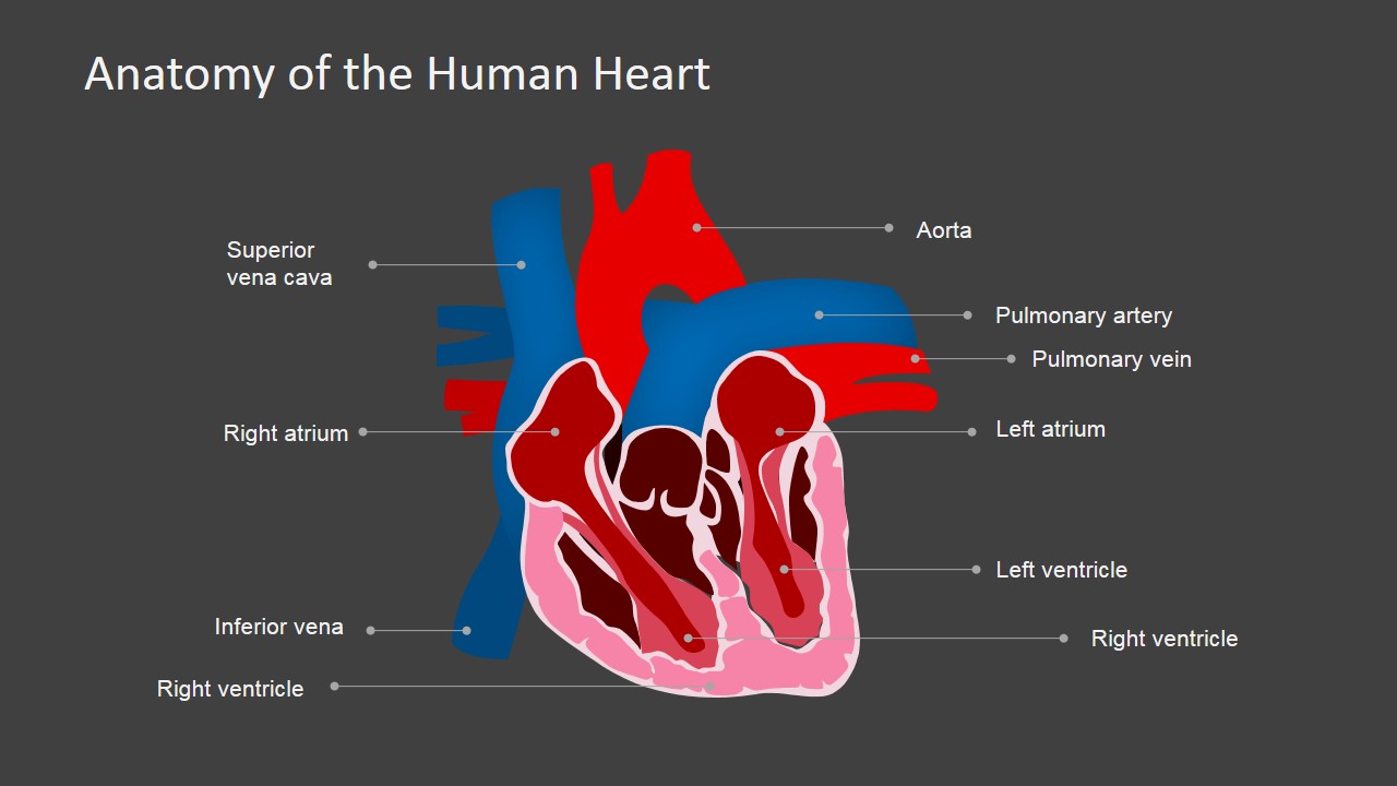
Anatomy of the Heart • The heart is composed of four chambers: • The right and left atria, which make up the top portion of the heart, are receiving chambers. • The right and left ventricles, which make up the bottom portion of the heart, are the discharging and pumping chambers.
Anatomy of the Human Heart PowerPoint Shapes SlideModel
PowerPoint Presentation Cardiac Anatomy & Physiology Circulatory System The heart is a hollow muscular organ made of specialised cells that allow it to act as a pump within the circulatory system Cardiovascular and lymphatic systems make up the circulatory system a vast network of organs and vessels responsible for the flow of:

Kardiyovasküler Sistem Anatomisi ve Fizyolojisi
Ads Heart: Anatomy and Function Presentation Premium Google Slides theme and PowerPoint template Present the fascinating world of the heart with our comprehensive presentation on its anatomy and function. This medical template features stunning heart illustrations, tables, graphs, and infographics to help you dive deep into the subject matter.

Pin on Nursing School and Study Guides
Heart anatomy. The heart has five surfaces: base (posterior), diaphragmatic (inferior), sternocostal (anterior), and left and right pulmonary surfaces. It also has several margins: right, left, superior, and inferior: The right margin is the small section of the right atrium that extends between the superior and inferior vena cava .